Mass transport studies in porous silica as a function of core-shell architecture
The study of molecular transport within the pores of silica is of great importance to many research areas; drug delivery, sensing, catalysis, filtration, encapsulation and gas sorption. Theoretical models describing transport behaviour have already been reported; however, few have presented a physical system for observing the accessibility of guest species in mesoporous silica. Here we report fluorescent core-shell particles as a platform to study molecular transport within a porous silica system with tuneable characteristics such as pore lengths, pore diameters and surface chemistry. The core particles were synthesised from non-porous silica spheres with post-synthetically grafted fluorescein moieties, Figure 1a. A silica shell was subsequently deposited around the functionalised cores. A variety of shell thicknesses were achievable through careful control of the addition rate of silane (tetraethyl orthosilicate (TEOS)) and hydrolysis solution. A primary shell (s1, approx. 800 nm, Figure 1b) demonstrated a very narrow particle size distribution which broadened on additional shell growth (s2, approx. 1300 nm, Figure 1c). The exterior of the core-shell particle was functionalised with rhodamine for convenient visualisation by CLSM, Figure 1d. The shell is inherently macroporous but well-defined mesopores can be introduced into the silica network through pseudomorphic transformation (PT), while retaining the original morphology. PT in addition to postsynthetic functionalisation generated a variety of pore sizes and pore surface chemistries. Core accessibility and surface wettability can then be determined by investigating the quenching of the fluorescent core.
The large variety of core-shell particles available through customisation enabled the testing of core accessibility as a function of the shell architecture.
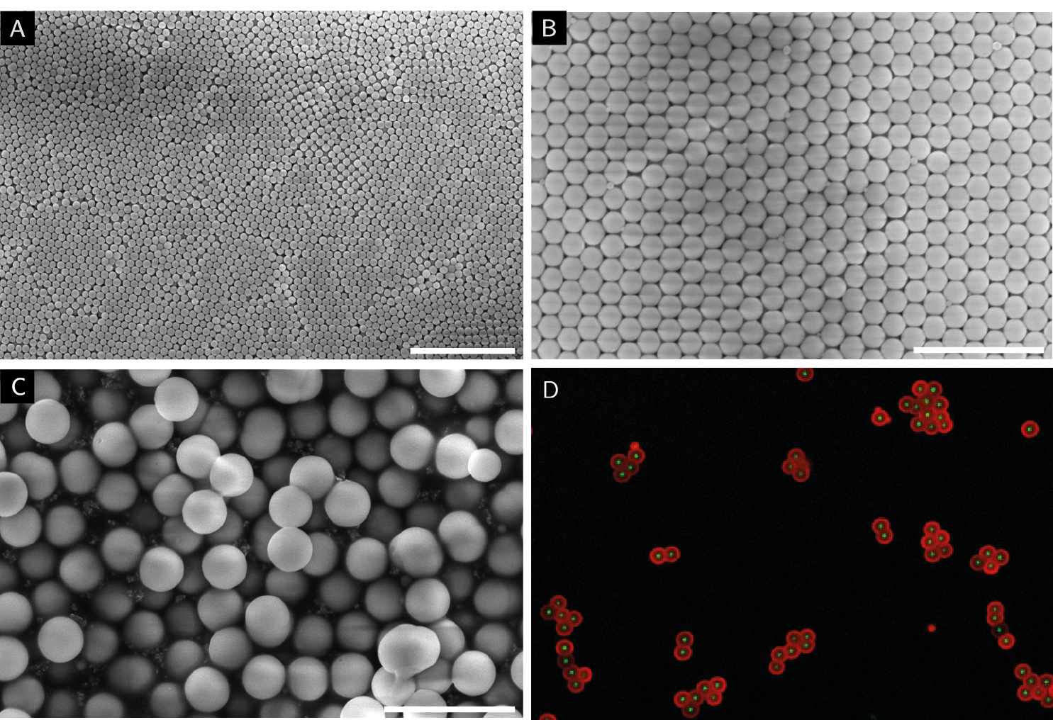
Figure 1) SEM images of the different stages of the functionalised core-shell particles. (A) Stöber particles, (B) core-shell (s1) particles, (C) core-shell (s2) particles and (D) a CLSM image of s2 particles functionalised with two dyes. Fluorescein on the core (green) and rhodamine B on the exterior (red). All scale bars are equal to 5 μm.